
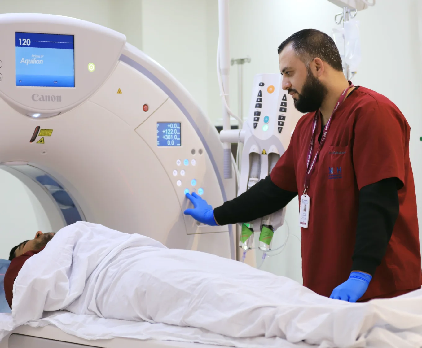
The department provides a wide range of diagnostic imaging services, with the appropriate imaging modality and protocol selected according to the clinical indication and diagnostic question. All diagnostic procedures can be performed under general anesthesia under the supervision of specialized physicians when required by the patient’s medical condition.
Routine and Specialized Services Include:
-
1.Magnetic Resonance Imaging (MRI):
The MRI system uses advanced digital technology and a multi-channel configuration for signal acquisition, resulting in high-quality, high-resolution images.
The system features a wide and comfortable bore, which can help reduce anxiety and discomfort in some patients.
The MRI system is capable of performing a wide range of specialized and advanced examinations, including:
- Cardiac MRI.
- Magnetic resonance enterography (MR Enterography).
- Magnetic resonance cholangiopancreatography (MRCP) for imaging the liver and biliary tree.
- MRI of the pancreas and pancreatic ducts.
- Magnetic resonance angiography and venography (MR Angiography and MR Venography), with or without contrast.
- MRI of the chest and abdominal organs.
- MRI of the brain and spine, including imaging of the Circle of Willis.
- MRI of all major joints and muscles, including MR arthrography with intra-articular contrast injection.
-
2. Computed Tomography (CT):
The department provides CT examinations, including contrast-enhanced studies, as well as multiplanar and three-dimensional reconstructions of various body regions and organs.
- Coronary CT angiography (Coronary CTA).
- CT angiography and venography of various body regions, including the upper and lower extremities, liver, kidneys, and brain.
- CT imaging of bones and joints.
-
3. Ultrasound and Doppler:
The department provides a wide range of ultrasound and Doppler examinations, including:
- Ultrasound examination of the abdominal and pelvic organs, including the liver, spleen, kidneys, urinary bladder, and uterus, as well as the thyroid gland.
- Doppler ultrasound of various organs and vascular structures, including the liver, kidneys, and arteries of the upper and lower extremities.
- Doppler ultrasound evaluation of arteriovenous dialysis access.
- Liver and portal vein Doppler ultrasound.
- Renal Doppler ultrasound.
- Ultrasound examination and follow-up of developmental dysplasia of the hip (DDH).
-
4. Digital and Emergency X-ray Imaging:
The department provides X-ray imaging of various body regions and the musculoskeletal system. The system features a long imaging field, allowing full-length imaging of the spine and single-image imaging of the entire limbs.
This capability is not widely available and is particularly useful for comprehensive skeletal imaging. The system also incorporates features designed to reduce patients’ radiation exposure and provides rapid image acquisition and processing.
- A dedicated X-ray system for outpatient clinics, designed to save patients time and effort.
- A range of portable X-ray units that can be transported to the patient’s location when transferring the patient is not feasible, such as in intensive care and emergency settings.
-
5. Fluoroscopy:
A modern digital fluoroscopy system that provides dynamic, real-time imaging and is used for a wide range of diagnostic examinations and image-guided procedures, including:
-
Esophageal, gastric, and intestinal imaging.
- Colon imaging.
- Hysterosalpingography (HSG) for imaging the uterus and fallopian tubes.
- Image-guided intra-articular injections.
- Other routine fluoroscopic examinations.
Doctors in this department

Dr. Laila Dmaidi
Breast Imaging and Interventional Radiologist
